By Lawrence Lin
GLGA 主席|GLG 董事会成员|IWBI WELL Light Concept Advisor|LRS 创始人兼 CEO
Today, while attending the Good Light Group (GLG) board meeting, I asked the directors—particularly our Chief Scientist, Marijke—about the significance of the newly announced Nobel Prize in Physiology or Medicine for photobiology research.
In our discussion, everyone agreed that this is an inspiring discovery for understanding the mechanisms connecting light and physiology.
For those of us who have worked in lighting for many years, the news deserves particular attention. Light does more than allow us to see objects; it can also participate in the transmission of biological signals. Science is increasingly providing answers to fundamental questions: Where does light act? Through what mechanisms? And what effects does it produce?
However, the more exciting a discovery is, the more important it is to describe it accurately.
This Nobel Prize was not awarded for a particular “healthy light,” nor does it demonstrate that everyday lighting can alter human genes. It recognizes the discovery of light-gated ion channels and optogenetics—a method that allows researchers to use light to precisely intervene in the activity of specific cells.[1]
1 | From 2017 to 2026: Two Different Scientific Paths
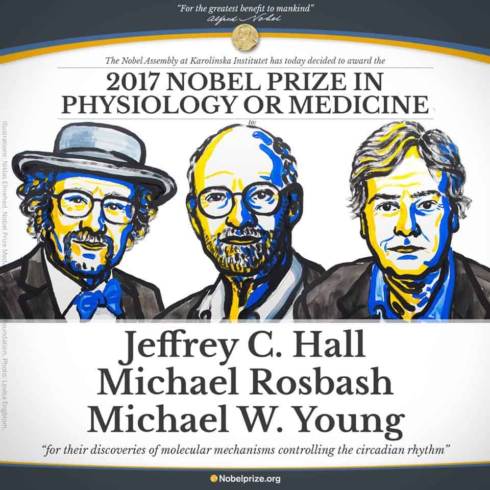
In 2017, Jeffrey C. Hall, Michael Rosbash, and Michael W. Young were awarded the Nobel Prize in Physiology or Medicine for their discoveries concerning the molecular mechanisms controlling circadian rhythms.
Their research revealed that the biological clock is not merely an abstract metaphor. Gene expression, protein accumulation, and feedback regulation within cells can form a continuously operating rhythmic mechanism that enables organisms to anticipate daily changes. These mechanisms are associated with physiological processes including sleep, hormone secretion, body temperature, and metabolism.
It is important to clarify that the 2017 prize recognized the molecular mechanisms of the biological clock. It was not awarded for the health effects of a particular lighting product, nor was it the first discovery that light affects the human body.
How light enters the circadian regulation system is part of another line of research. A key study published in 2002 identified intrinsically photosensitive retinal ganglion cells and their connection to circadian regulation. This provided an important foundation for subsequent understanding of melanopsin, ipRGCs, and the non-visual effects of light.
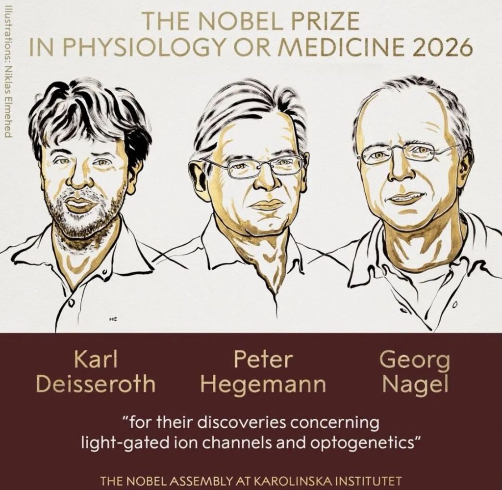
In 2026, Karl Deisseroth, Peter Hegemann, and Georg Nagel were recognized for showing another possibility: using light-sensitive proteins to turn specific cells into targets that can receive optical commands.[1]
The two Nobel Prizes are not successive confirmations of the same mechanism.
One revealed how life keeps time; the other developed a tool for controlling cellular activity with light. For light research, together they broaden our understanding of how light can interact with living systems.
2 | Optogenetics: Turning Light into a Cellular Control Signal
The key to this research came from light-sensitive proteins found in microorganisms.
Take channelrhodopsin-2 (ChR2) from green algae as an example. It can both sense light and function as an ion channel in the cell membrane. Research published in 2003 demonstrated that when ChR2 is expressed in animal cells, light can directly open the channel, allowing cations to flow across the membrane.
Researchers subsequently introduced genes encoding these proteins into neurons. Cells that originally lacked this light-sensing capability thereby acquired an optical control interface.
Light-sensitive protein absorbs a photon → protein changes conformation → channel opens → ions flow → membrane potential changes → the neuron may fire.
The actual photon absorber is the retinal chromophore within the protein. After absorbing light, it undergoes isomerization, driving the protein into a different photochemical state and changing the conductance of the channel.
Under typical resting conditions in a neuron, opening ChR2 produces a net inward cation current, depolarizing the cell. Once a sufficient threshold is reached, the neuron’s own voltage-gated channels generate an action potential.
Light is the triggering signal here. Neural firing still depends on the cell’s existing ion gradients, electrophysiological state, and channel systems.
In 2005, a landmark study by Boyden, Deisseroth, Nagel, and colleagues demonstrated the ability to control neuronal firing on a millisecond timescale using brief pulses of light.[2] Researchers subsequently developed light-driven inhibitory tools, meaning that light could be used not only to activate neural activity but also to suppress it.
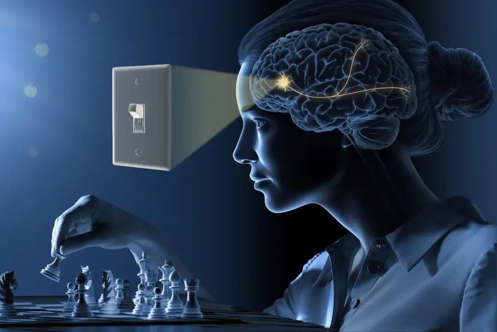
© The Nobel Committee for Physiology or Medicine. Ill. Mattias Karlén
3 | Why Is This a Major Breakthrough in Research Methodology?
Observing that certain neurons become active during a particular behavior does not mean that we know those neurons cause the behavior.
They could be the cause, the consequence, or simply be involved in another process occurring at the same time.
Optogenetics enables researchers to intervene directly: activate a particular class of cells and observe whether a response occurs; inhibit them and see whether the response is reduced; change the timing of stimulation and examine how the outcome changes.
This gives neuroscience a much stronger ability to test causality rather than stopping at observations of correlation.
Of course, a single intervention cannot automatically explain all the causes of a complex behavior. The accuracy of cell targeting, possible unintended effects of optical stimulation, and the adequacy of experimental controls all remain critical to the reliability of the conclusions.
For us, this methodology is particularly worth learning from: research into light and health requires clearly defined intervention conditions, as well as experimental designs capable of ruling out alternative explanations.
4 | It Is Not a “Magic Wavelength” That Makes the Difference
Early ChR2 studies commonly used blue light around 470 nm. The 2005 experiment used a 450–490 nm range and compared short stimulation pulses of 5, 10, and 15 milliseconds.[2]
Different light-sensitive tools developed later can operate at different wavelengths. For example, the light-driven chloride pump NpHR uses yellow light to inhibit neural activity, while ChrimsonR, used in some visual-restoration research, is stimulated with orange light around 595 nm.
This does not mean that blue light is inherently associated with “excitation,” or that orange light is inherently associated with “vision restoration.”
The effect depends on which protein is expressed by the target cell and whether the light can effectively reach the target.
Likewise, dose cannot be adequately described using a single total-energy value. At a minimum, we need to specify:
- Wavelength and spectral distribution;
- Irradiance or photon flux at the target cells;
- Pulse duration, intervals, and stimulation sequence;
- Expression level, activation/deactivation kinetics, and recovery state of the photosensitive protein;
- Absorption and scattering of light within the tissue.
The photocurrent generated by ChR2 exhibits a peak, followed by decay and recovery. Continuous illumination and short pulses can produce different firing patterns even when the total exposure is the same. Subsequent models of the photocycle were developed precisely to describe these dynamic changes.
Therefore, wavelength is only the starting point. Where the light reaches the tissue, and the temporal pattern in which it arrives, can be equally important.
5 | From Research Tool to Medical Application
Optogenetics has now entered human research.
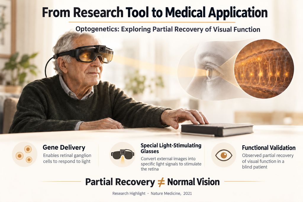
In 2021, Nature Medicine reported partial recovery of visual function in a blind patient. Researchers delivered a vector encoding ChrimsonR into the eye and used specialized goggles to convert visual information into light pulses projected onto the retina.
After treatment, and while wearing the goggles, the patient was able to perform certain tasks involving object perception, localization, counting, and reaching. Without the goggles, the patient did not demonstrate the same ability to detect objects.[3]
This was an important clinical exploration, but its implications have clear boundaries: it did not restore normal vision, and it was not an effect produced by ordinary lighting.
The promise came from a specific biological target, gene delivery, an optical system, and functional validation. None of these components can simply be omitted.
6 | The Lighting Industry: Moving from “What Light Is There?” to “What Light Does a Person Actually Receive?”
Everyday lighting and optogenetics operate through different mechanisms. However, the precise description of stimulation conditions in optogenetics provides valuable inspiration for lighting research.
We are accustomed to describing light from the product side: power, luminous flux, correlated color temperature, color rendering, and spectrum.
When the question concerns physiological responses in people, we also need to know: after light passes through a space, reflection, obstruction, and patterns of use, how much actually reaches the eye, when does it arrive, and for how long?
This is the shift from source specifications to actual human exposure.
CIE S 026 establishes a measurement framework for describing the stimulation of five classes of photoreceptors in the human eye. Mel-EDI is one of the metrics used to describe melanopsin-related stimulation, but it is not a direct measure of sleep quality, cognitive performance, or overall health outcomes.
A field measurement can tell us whether a particular stimulation condition has been achieved. Determining the resulting human outcome requires the corresponding research evidence.
Likewise, photobiomodulation research involving red or near-infrared light should not borrow the mechanism of optogenetics as evidence of efficacy. Different pathways require their own exposure models, dose-response relationships, and evidence of effects.
6 | What Should Future Light Research Prioritize?
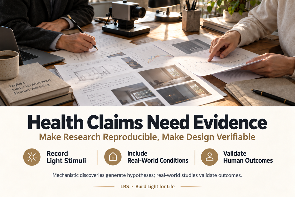
From the LRS perspective, we believe three areas deserve continued attention.
01. Make Research Conditions Reproducible
Without clear information about the spectrum, measurement location, timing protocol, and equipment, research findings are difficult to compare and even more difficult to translate into reproducible applications.
CIE TN 016:2026 proposes the ENLIGHT checklist for reporting optical characteristics in laboratory-based human light-intervention studies, with the aim of improving research reproducibility and comparability. It is not a standard specifically for optogenetics, but it reflects the same fundamental requirement: the light stimulus must be described clearly and completely.
02. Include the Space and the User in Design and Validation
The same luminaire can produce different ocular exposure depending on the space, viewing direction, and time of use. Research and design should not stop at the light source. They should also establish the connection between spatial models, actual measurements, and records of real-world operation.
03. Match Every Efficacy Claim to Its Evidence
“More comfortable,” “supports circadian regulation,” “improves sleep,” and “enhances learning performance” are not interchangeable claims. Each outcome requires an appropriate study population, measurement method, and control condition.
Mechanistic discoveries can help us formulate better hypotheses; product efficacy still has to be answered by evidence concerning the product and the context in which it is used.
8 | LRS: Build Light for Life
Today’s board discussion left me feeling both inspired and responsible.
Inspired because light research is continually expanding our understanding of life.
Responsible because the industry must understand these discoveries accurately—neither overlooking them nor turning them into claims that go beyond the evidence.
光配方研究院(Lighting Recipe Studio, LRS) focuses on research into healthy lighting environments, sensing technology, testing and monitoring equipment, algorithm capabilities, and spatial application systems. We care not only about lighting outcomes, but also about the true relationships between light, people, space, time, and activities. Our aim is to explain important research clearly, distinguish between different mechanisms, and strengthen the connection between design, measurement, and real-world validation.
What the Nobel Prize gives us is not a health label that can simply be attached to a luminaire.
It shows us that patiently pursuing a fundamental question can ultimately lead to discoveries that transform the way research is conducted.
The relationship between light and health involves mechanisms that can be studied and outcomes that can be tested. The value created by the lighting industry in its next stage will depend on whether we can turn this knowledge into designs and applications that stand up to rigorous validation.
Light and health are not pseudoscience.
Ensuring that every health claim can be traced back to evidence is work we all share
— LRS
Further Reading and Original Sources
[1] Official 2026 Nobel Prize in Physiology or Medicine Announcement
Award topic: The discovery of light-gated ion channels and optogenetics.
https://www.nobelprize.org/prizes/medicine/2026/press-release
[2] Landmark 2005 Optogenetics Paper
Boyden ES, Zhang F, Bamberg E, Nagel G, Deisseroth K.
Millisecond-timescale, genetically targeted optical control of neural activity.
Nature Neuroscience, 8, 1263–1268 (2005).
The study demonstrated the use of ChR2 expression to control neuronal activity on a millisecond timescale using pulses of light.
https://doi.org/10.1038/nn1525
[3] 2021 Study on Partial Recovery of Visual Function in Humans
Sahel JA et al.
Partial recovery of visual function in a blind patient after optogenetic therapy.
Nature Medicine, 27, 1223–1229 (2021).
The study combined gene delivery with specialized light-stimulation goggles and reported partial recovery of visual function in a blind patient. The effect was not produced by ordinary lighting.
请注意: The board discussion was compiled from the author’s meeting notes. The scientific interpretation and industry recommendations in this article were written by the author and do not constitute direct quotations from individual scientists or an official position statement of GLG. The cover image is a conceptual visual creation and does not represent an experimental apparatus or clinical treatment setting.

 English
  Chinese (Taiwan)
  Chinese (China)
  German